GC-MS
How Does Radiation Affect Gastrointestinal Function? - Chromatography Investigates
Sep 01 2020
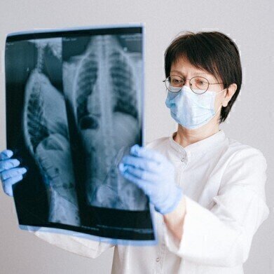
Radiotherapy is a treatment that is widely used in cancer treatment. According to a paper published by researchers in Japan approximately half of cancer patients undergo radiation therapy and the treatment plays a role in curing almost half of those cases. But unfortunately, radiation therapy can have some side effects.
In a paper published in the journal Biochemistry and Biophysics Reports, researchers in Japan have been investigating the changes in the metabolic profile after irradiation of the intestine. Irradiation of pelvic tumours can damage nearby tissues in the intestine and the researchers were interested in the intestinal-specific metabolic markers after radiation exposure. The results were published in a paper - Elucidation of gastrointestinal dysfunction in response to irradiation using metabolomics – and chromatography played a role.
What is radiotherapy?
Put simply, radiotherapy is a treatment that uses radiation to kill cancer cells. It can be used in the early stages of cancer or when cancer cells have started to spread around the body. It is used to cure cancer completely, or to make other treatments more effective – a combination of radiotherapy and chemotherapy is known as concurrent therapy and is sometimes used when surgery is not an option. Radiotherapy is one of the most effective cancer treatments after surgery, but its effectiveness varies between people.
Radiotherapy can be administered in several ways. A machine can be used to direct beams of radiation of the site of the cancer; or a small piece of radioactive metal can be implanted inside the body near the cancer; or radioisotope therapy can be used where a radioactive liquid is swallowed or injected into the bloodstream. But, as mentioned above, radiation can kill healthy cells along with the cancer cells. Most of the side effects cease when the treatment stops. But the team of researchers in Japan investigated the effect of radiation therapy after treatment for pelvic malignancies.
Chromatography helping with radiation treatment
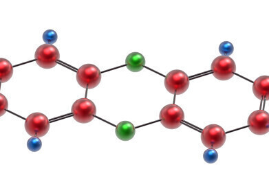
The primary goal of the study was to identify the metabolic markers that were changed during abdominal exposure to X-ray radiation at 2Gy and 20Gy doses. They used mice as the subjects, and they were exposed to x-ray radiation after being anesthetised. After treatment, intestinal samples were taken and the abundance of metabolites in the intestine were analysed using gas chromatography- mass spectrometry. The use of GC-MS is discussed in the article, Analysis and Identification of Mezcal and Tequila Aromas by Ambient Ionisation MS, GC-MS, and GCxGC-MS.
The researchers detected 44 metabolites in both control and radiation groups that they classified as carbohydrate, lipid, and protein intermediates. The study showed that there was an increase in amino acids in the intestinal tissue after radiation. The team are unsure why the amino acid levels increased and how the increased levels protect against radiation. The amino acids could affect the normal response to stress and damage and could be a marker for gastrointestinal toxicity due to radiation exposure.
Digital Edition
Chromatography Today - Buyers' Guide 2022
October 2023
In This Edition Modern & Practical Applications - Accelerating ADC Development with Mass Spectrometry - Implementing High-Resolution Ion Mobility into Peptide Mapping Workflows Chromatogr...
View all digital editions
Events
Jan 20 2025 Amsterdam, Netherlands
Feb 03 2025 Dubai, UAE
Feb 05 2025 Guangzhou, China
Mar 01 2025 Boston, MA, USA
Mar 04 2025 Berlin, Germany


