Electrophoretic separations
Identification process of cancer cells is now safer and simpler
Oct 26 2012
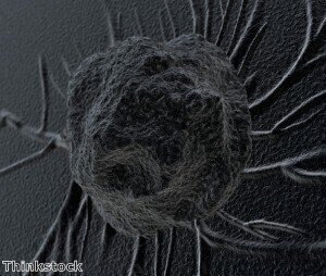
A new fluorescent glucosamine probe can make identification of cancer cells using two-photon microscopy simpler and safer.
Typically, the early discovery of soft-tissue diseases, such as breast cancer, needs invasive biopsies.
A new self-assembled nanoparticle developed by Bin Liu and a team from the A*STAR Institute of Materials Research and Engineering might soon make biopsies redundant.
The safety of two-photon microscopy (TPM) has considerably increased due to the creation of the team's material, called dendrimer.
Even though TPM offers deep access to cell tissue without causing serious photo-damage, the challenging issue is finding appropriate substances to act as light-emitting probes.
Exceptional cell-structure illuminators named "quantum dots" are nanoscale collections of elements such as selenium and cadmium.
These are excellent illuminators due to their stable and bright fluorescence, however biological applications are restricted because of their innate toxicity.
Conjugated organic molecules were then utilised to produce less toxic dyes for TPM by Ms Liu and her team.
The problem with dealing with such tiny organic molecules is that they are usually unable to absorb enough amounts of laser light to begin fluorescence imaging, but the team has synthesised a star-shaped material known as dendrimer to solve this problem.
Dendrimer has a central triphenyl amine core and three 'arms' made from extended conjugated chains.
This distinctive geometry can induce bigger cross sections that can absorb two-photons better than isolated fluorescent dyes.
Researchers had to use a chemical trick to make sure there is biocompatibility between the star-shaped dendrimer and cell tissue.
Using a mild bromide–thiol reaction, the team attached several glucosamine sugar rings to the dendrimer's arms.
This method decreased the cytotoxicity of the dye and allowed them to functionalise it with folic acid ligands that target the surfaces of MCF-7, which is a breast cancer cell line.
When submerged in water, the experiments showed that the dendritic dye self-assembled into dispersed nanoparticles. This form gives a high yield of laser-induced fluorescence and increases two-photon-absorption cross sections.
Subsequent TPM imaging showed a bright fluorescence localised inside the cancer cell cytoplasma following the incubation of these nanoparticles into the MCF-7 cells.
Specific binding occurs between dendritic dye and folate receptors on the MCF-7 surface.
Confirming that this strategy is a safe and easy way to increase the use of TPM imaging are the presence of cell viabilities close to 100 per cent at dye concentrations.
"We are keen to expand the current in vitro imaging to in vivo applications," said Ms Liu.
Posted by Ben Evans
Events
Feb 03 2025 Dubai, UAE
Feb 05 2025 Guangzhou, China
Mar 01 2025 Boston, MA, USA
Mar 04 2025 Berlin, Germany
Mar 18 2025 Beijing, China


